Annals of Environmental Science and Toxicology
Neurotoxicity of Zero-Valent Iron Nanoparticles in Primary Zebrafish Brain Cell Cultures
1Departamento de Ciencias Biologicas, 12 Ave. Universidad STE. 1201, Universidad de Puerto Rico, Recinto de Rio Piedras, San Juan, 00925-2532, USA
2School of Science & Technology, Ana G. Mendez University, USA
Author and article information
Cite this as
Hidalgo KS, et al. Neurotoxicity of Zero-Valent Iron Nanoparticles in Primary Zebrafish Brain Cell Cultures. Ann Environ Sci Toxicol. 2026; 10(1): 41-48. Available from: 10.17352/aest.000095
Copyright License
© 2026 Hidalgo KS, et al. This is an open-access article distributed under the terms of the Creative Commons Attribution License, which permits unrestricted use, distribution, and reproduction in any medium, provided the original author and source are credited.Zero-valent iron nanoparticles (nZVI) are increasingly used in environmental remediation and explored for biomedical applications; however, their potential neurotoxic effects and underlying cellular mechanisms remain incompletely understood. In this study, newly synthesized nZVI were physicochemically characterized and evaluated for cytotoxic effects using primary brain cell cultures derived from adult zebrafish (Danio rerio) as an in vitro model of neurotoxicity. Nanoparticles were characterized by scanning electron microscopy (SEM), which revealed spherical morphologies with aggregate chain formation, and by X-ray photoelectron spectroscopy (XPS), confirming the presence of mixed metallic and oxidized iron states at the surface. Primary zebrafish brain cells were exposed to increasing concentrations of nZVI for 48 hours to assess cell viability, apoptotic cell death, and mitochondrial membrane integrity. Cell viability assays demonstrated a concentration-dependent decrease in metabolic activity, with a 50% growth inhibition (GI₅₀) observed at approximately 220 ng/mL. Apoptosis analysis using Annexin V/propidium iodide staining showed limited and non-significant activation of apoptotic pathways across the tested concentrations. In contrast, mitochondrial membrane damage assays revealed significant, dose-dependent mitochondrial dysfunction beginning at 188 ng/mL. Together, these findings indicate that nZVI induces cytotoxic effects in primary zebrafish brain cells that are closely associated with mitochondrial dysfunction and occur largely independent of classical apoptotic cell death. This in vitro model informs the risk assessment and safe development of iron-based nanomaterials for environmental and biomedical research contexts.
Heavy metal exposure has been linked to negative health effects, including respiratory disorders, cardiovascular disease, and neurodegenerative conditions [1,2]. At the cellular level, metal-induced toxicity often involves oxidative stress, mitochondrial dysfunction, and disrupted redox balance, especially in neural tissues that are highly vulnerable to iron buildup and reactive oxygen species production [3,4]. Nanoscale zero-valent iron nanoparticles (nZVI) are a new class of materials used for environmental cleanup [5,6]. Additionally, nZVI have been extensively studied for biomedical applications because of their high reactivity and magnetic properties [7,8]. However, growing evidence suggests that nZVI can undergo physicochemical changes in biological environments, such as aggregation, surface oxidation, and protein corona formation, which can greatly affect their biological interactions and toxicity profiles [9-11].�Despite considerable research on iron-based nanomaterials, the cellular mechanisms behind their neurotoxic effects in primary neural cell cultures are still not fully understood, especially regarding mitochondrial responses and non-apoptotic pathways [12,13]. Most studies have focused on whole-organism models or non-neural cell lines, leaving key gaps in our knowledge of mitochondrial responses, oxidative stress pathways, and non-apoptotic injury mechanisms caused by nZVI [13-15]. Many studies promoting nZVI use tend to highlight its benefits, with few investigating its toxicity mechanisms, uptake, translocation, or bioaccumulation in organisms, including plants and animals within ecosystems [16]. Zebrafish (Danio rerio) have become a valuable model in toxicology research because of their genetic similarity to humans, well-understood nervous system, and suitability for both in vivo and in vitro studies [17-19]. Primary zebrafish brain cell cultures offer a biologically relevant platform for mechanistic neurotoxicity testing while enabling controlled exposure and direct evaluation of cellular responses [20-22]. They are an excellent model for conducting in vitro experiments and developmental toxicology studies, helping us understand the mechanisms behind various human conditions. According to a study by the Sanger Institute in the United Kingdom, sequencing the zebrafish genome shows a high level of conservation with humans, with about 70% of their protein-coding genes and 84% homology [17].
Accordingly, this study aimed to (i) characterize newly synthesized zero-valent iron nanoparticles using scanning electron microscopy and X-ray photoelectron spectroscopy, and (ii) evaluate their cytotoxic effects in primary zebrafish brain cell cultures by quantifying dose-dependent changes in cell viability, apoptotic signaling, and mitochondrial membrane integrity following 48 hours of exposure. However, the cellular mechanisms underlying nZVI-induced neurotoxicity in primary zebrafish brain cells remain insufficiently characterized.
Materials and methods
Synthesis of zero-valent iron nanoparticles
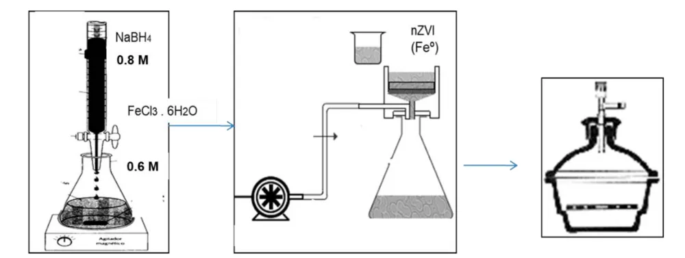
Zero-valent iron nanoparticles (nZVI) were synthesized by reducing ferric chloride hexahydrate (FeCl36H2O) with sodium borohydride (NaBH4). A 0.6 M FeCl36H2O solution was prepared using an ethanol (83%v/v)-Nanopure water mixture; the supplementary protocol specifies 30 mL of ethanol but does not establish an unambiguous final solution volume. The iron precursor solution was purged with nitrogen for 30 minutes. Under a nitrogen atmosphere, 100 mL of 0.8 M aqueous NaBH4 was added. The reaction mixture was stirred magnetically at 700 rpm for 30 minutes. The solid was recovered by vacuum filtration through a 0.22 �m Millipore membrane at approximately 25�C, washed three times with 99.5% absolute ethanol, and immediately transferred to a vacuum desiccator. For cell exposures, 0.003 mg of recovered nZVI was dispersed in 1 mL of dimethyl sulfoxide (DMSO), producing a nominal 3�g/mL suspension. These concentrations represent nominal added nZVI mass; nanoparticle aggregation, dissolved iron, and delivered cellular dose were not measured (Figure 1).
2 Fe3+ + 6 BH4- + 18 H2O ? 2 Fe0(s) + 6 B(OH)3 + 21 H2
Zebrafish husbandry and primary brain cell culture preparation
Adult zebrafish (Danio rerio) were obtained from Caribe Fisheries Inc., a tropical aquaculture farm in Lajas, Puerto Rico, and transported to the ChEMTox Laboratory at UAGM-Cupey. Upon arrival, fish were acclimated to a tank containing 5 L of distilled water and 5 L of tap water in a 20 L aquarium. The transport bag was placed in the tank for 15 minutes before the fish were released. Water temperature was maintained at 26�C, water was changed every two weeks, and fish were maintained for at least 24 hours before experimentation. Euthanasia procedures followed the American Veterinary Medical Association's 2013 Guidelines for the Euthanasia of Animals; ARRIVE reporting principles were also considered.
Primary brain cell cultures were prepared from adult zebrafish using a dissection protocol adapted from Carrasquillo et al. (2021) (Figure 2). Fish were immersed in cold water to reduce mobility and immediately decapitated on a chilled dissection surface. Heads were immersed briefly (2-5 seconds) in 75% ethanol, and brains were isolated in dissection medium. The supplementary protocol describes DMEM/F12 supplemented with penicillin/streptomycin for dissection and subsequent transfer of isolated brains to Neurobasal medium supplemented with Primocin (10 �L per 5 mL Neurobasal). Brain tissue was mechanically dissociated to obtain primary cell cultures. Cultures were allowed to stabilize for 24 hours before nanoparticle exposure. For the PrestoBlue assay, cells were cultured in 96-well plates with 200 �L of Neurobasal medium per well. For toxicity assessment, primary neural cells were exposed to increasing concentrations of nZVI (0-375 ng/mL) for 48 hours.
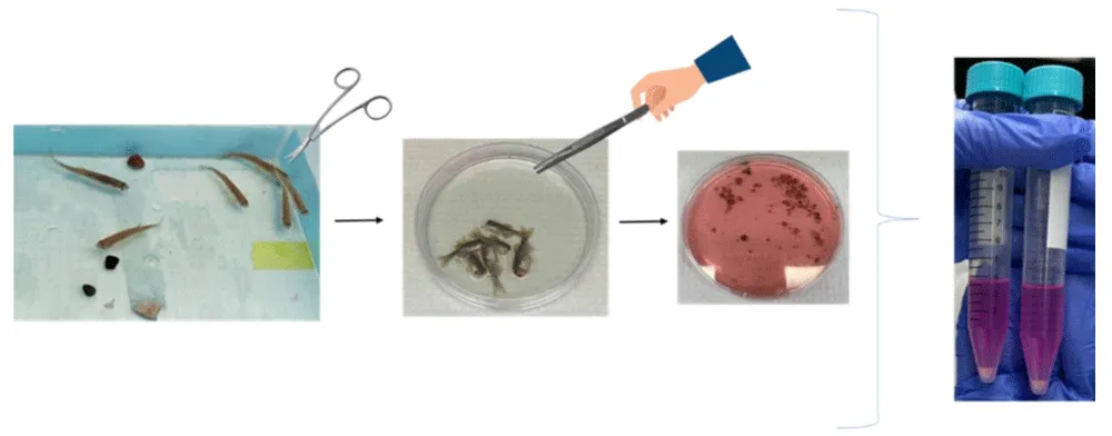
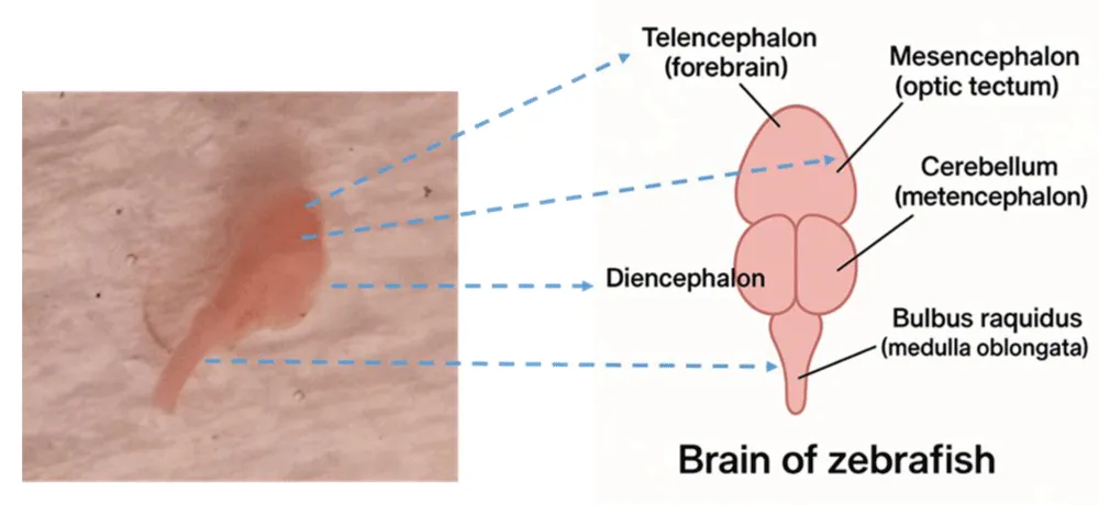
This procedure protects the brain from infection, maintains its functional and structural viability, and prepares it for more advanced studies (Figure 3).
Measures of variables
Nanoparticle characterization was performed using scanning electron microscopy (SEM) to assess particle morphology and aggregation behavior, and X-ray photoelectron spectroscopy (XPS) to determine surface chemical composition and oxidation states. Cell viability was evaluated using the PrestoBlue metabolic assay after 48 hours of exposure, with fluorescence intensity used as an indicator of cellular metabolic activity. Apoptotic cell death was assessed using Annexin V and propidium iodide staining, which allowed for the identification of apoptotic and non-apoptotic cell populations. Mitochondrial membrane integrity was evaluated using the ToxGloTM mitochondrial toxicity assay as an indicator of mitochondrial dysfunction.
Data analysis procedure
All experiments were conducted in triplicate. Fluorescence data were collected using a FLUOstar OMEGA microplate reader.
The GI50 value was calculated using nonlinear dose-response curve fitting. Statistical significance was assessed using one-way analysis of variance (ANOVA) with multiple comparisons, performed using GraphPad Prism (version 9). Statistical significance was set at p < 0.05.
Results and discussion
Characterization of nZVI particles
Characterization of zero-valent iron nanoparticles is essential for understanding their reaction mechanism and kinetics. The transport, distribution, and fate of nanoparticles in the environment and within organisms also depend on these surface properties.
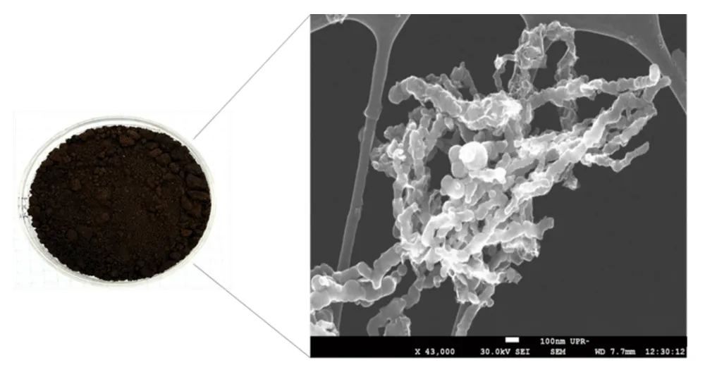
Scanning electron microscopy (SEM) was employed to examine the morphology and aggregation behavior of the synthesized zero-valent iron nanoparticles. SEM images were collected at a magnification of 43,000x with an accelerating voltage of 30.0 kV under high-vacuum conditions. Samples were analyzed in the dry state without dispersion in liquid media. Figure 4 shows the obtained micrographs that reveal nanoscale primary particles forming larger aggregated and chain-like structures, a characteristic feature of iron-based nanoparticles driven by magnetic interactions [23,24]. Particle size estimation was based on direct measurements of clearly resolved primary particles using the scale bar provided in the micrographs. SEM images revealed that the synthesized nZVI exhibit predominantly spherical primary particle morphologies with a strong tendency to form aggregated chain-like structures [25,26]. Particle size estimates were derived from direct measurements of clearly resolved primary particles in SEM micrographs under dry conditions; however, no hydrodynamic diameter or zeta potential measurements were performed, which represents a limitation of this characterization approach. While primary particle dimensions fall within the nanoscale range, aggregation results in the formation of larger secondary structures. The aggregation behavior observed by SEM reflects the intrinsic magnetic interactions of zero-valent iron nanoparticles and is consistent with previous reports describing nanoscale iron particle assembly and surface oxidation [24,25]. In biological environments, such physicochemical transformations, including protein corona formation, can substantially modify nanoparticle bioavailability and toxicity, underscoring the importance of contextualizing in vitro findings within dynamic exposure conditions.
Nanoparticles were also characterized using X-ray photoelectron spectroscopy (XPS). XPS binding energy spectra were obtained using a PHI spectrometer equipped with an Al Ka mono- and polychromatic X-ray source operating at 15 kV, 350 W, and a pass energy of 58.70 eV. All the binding energies reported were corrected, fixing the carbon 1s peak (C 1s) at 284.5 eV. XPS was used to characterize the surface chemical composition and oxidation states of the synthesized nZVI. Deconvolution of the Fe 2p3/2 spectrum revealed a dominant peak at approximately 711 eV, characteristic of ferric iron (Fe3+), along with a lower binding energy component near 707 eV corresponding to metallic iron (Feo) (Figure 5). This indicates partial surface oxidation of the nanoparticles while preserving a metallic iron core. Analysis of the O 1s region revealed multiple components at binding energies of approximately 529.9 eV, 531.2 eV, and 532.5 eV, consistent with lattice oxygen (O2-), hydroxyl groups (OH-), and adsorbed water species, respectively (Figure 6). The relative peak area ratios suggest that the surface oxide layer is composed primarily of iron oxides and oxyhydroxides. The coexistence of metallic iron (Feo) and oxidized iron species at the nanoparticle surface is relevant to their redox reactivity and may influence biological interactions through iron ion release and oxidative processes, potentially contributing to the cellular responses observed in subsequent toxicity assays [27-29]. This study presents an integrated evaluation of the neurotoxic effects of zero-valent iron nanoparticles in primary zebrafish brain cells, combining physicochemical characterization with functional cellular assays. By jointly assessing viability, apoptotic activation, and mitochondrial integrity, the present work identifies mitochondrial dysfunction as an early and central event in nZVI-induced cytotoxicity. Previous studies on iron-based nanoparticles have predominantly focused on whole-organism models, environmental remediation contexts, or cancer cell lines, often emphasizing oxidative stress and apoptotic outcomes [30,31]. In contrast, the present study employs primary neural cultures derived from adult zebrafish, a biologically relevant in vitro model that preserves key neuronal characteristics while allowing controlled mechanistic interrogation [20,21]. The observed dissociation between mitochondrial dysfunction and apoptotic activation adds to growing evidence that iron-mediated neurotoxicity may proceed through non-apoptotic mechanisms, particularly under conditions of direct cellular exposure [32,33]. Compared to previous whole-organism zebrafish studies, this in vitro neural model isolates direct cellular responses to iron exposure [18,19].
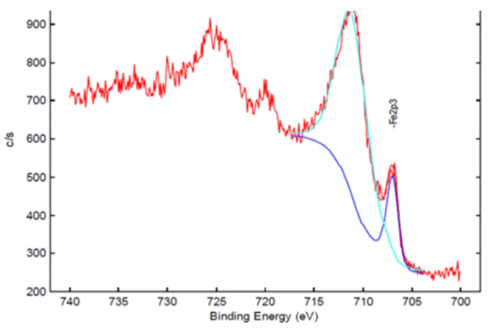
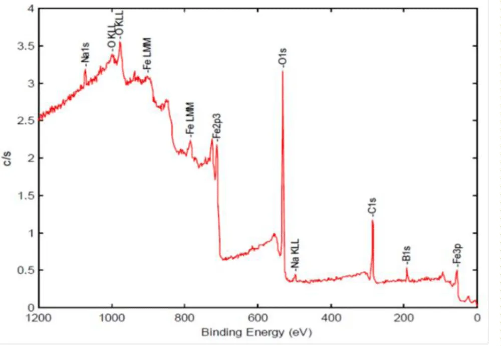
A summary of the key physicochemical and biological findings, together with their proposed mechanistic interpretation, is provided in Table 1. Taken together, the physicochemical characterization and cellular assays reveal a consistent pattern in which nZVI surface properties, mitochondrial dysfunction, and loss of metabolic activity are closely linked.
GI50 determination
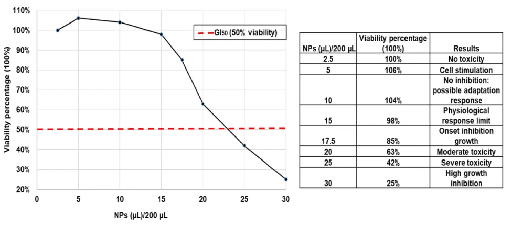
The GI50 value was determined by analyzing data with the MARS software on the FLUOstar OMEGA reader, using optical curve fitting to calculate the effective dose that inhibits cell growth by 50%. Figure 7 shows the dose-dependent effects of iron nanoparticles on primary zebrafish brain cells after 48 hours of exposure.
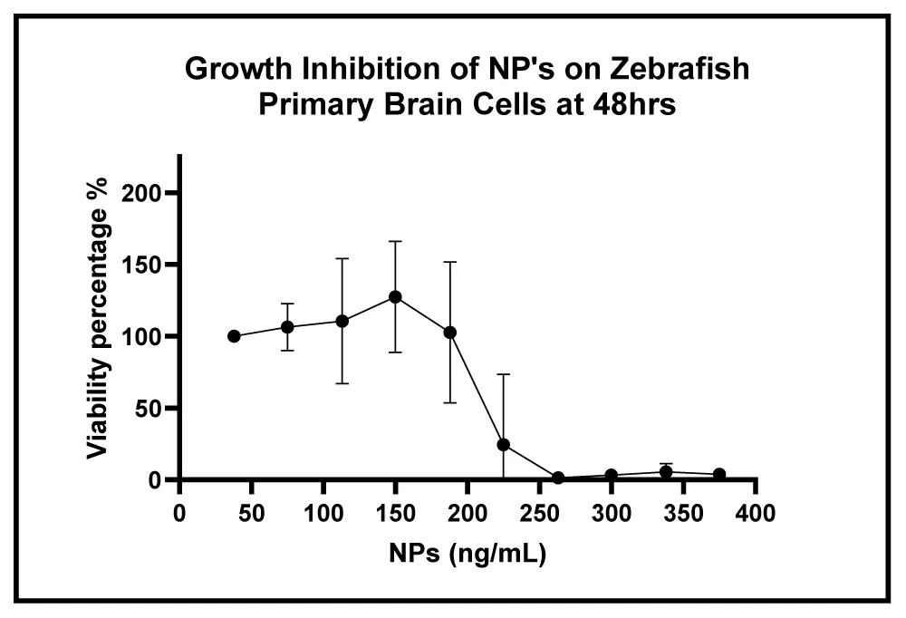
The control group (2.5 �L), composed of Neurobasal medium and brain cells without exposure to nanoparticles, maintained 100% viability, with no signs of growth inhibition [13,20]. Exposure of primary zebrafish brain cells to increasing concentrations of nZVI resulted in a dose-dependent reduction in metabolic activity, with a calculated GI50 of approximately 220 ng/mL. Similar concentration-dependent cytotoxic effects of iron-based nanoparticles have been attributed to mechanisms involving the generation of reactive oxygen species, mitochondrial impairment, and disruption of intracellular iron homeostasis [3,12,13,31]. These results are fundamental, since they confirm that the basal conditions of the assay do not induce toxicity or affect cell proliferation, which is consistent with studies that have established that the conditions of the culture medium critically influence the stabilization of the primary culture model [20,21]. In subsequent columns, corresponding to exposures to low concentrations of nanoparticles, an increase in cell viability was observed (=100%). This phenomenon may be attributed to a possible adaptive or proliferative response induced by nanoparticles at sub-cytotoxic concentrations. Such behavior has been documented in other biological models, where mild exposure to metal nanoparticles stimulates signaling pathways associated with proliferation [16]. Starting at a dose of 17.5 �L, a progressive decrease in cell viability was observed, indicative of growth inhibition. At this dose, the concentration of nZVI is 0.0525 �g. This point represents a threshold of toxicity and suggests a dose-dependent effect of nanoparticles on brain cells [13-15].
Figure 8 shows the cell growth inhibition curve along with the interpretation of the data obtained. The dashed red line indicates the GI50 point, which helps visualize the concentration at which the nanoparticles begin to have a significant cytotoxic effect. Several studies have shown that iron nanoparticles, at high concentrations, can induce neuronal toxicity through mechanisms such as the generation of reactive oxygen species (ROS), mitochondrial damage, and disruption of iron homeostasis, processes that can culminate in multiple forms of cell injury and death, depending on cellular context [13,14,31]. The reduction in cell viability observed at higher nZVI concentrations reflects a loss of metabolic activity rather than selective activation of programmed cell death pathways. The PrestoBlue assay primarily measures mitochondrial metabolic function; therefore, decreases in signal intensity are indicative of impaired cellular respiration and redox activity [13,26]. This interpretation is supported by the mitochondrial membrane damage detected at lower concentrations, suggesting that mitochondrial dysfunction precedes overt loss of cell viability [13,14,31].
Apoptosis-annexin V assay
Figure 9 depicts the apoptotic response of primary Danio rerio brain cells after 48 hours of exposure to different concentrations of iron nanoparticles. For this study, the percentage of apoptotic cells was compared with a negative control (without treatment) and a positive control treated with cisplatin, a chemotherapeutic agent known to induce apoptosis [17,27].
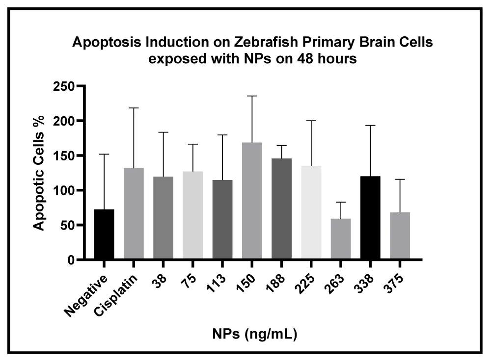
The results show that apoptosis, as the mechanism of cell death at the tested nanoparticle concentrations (220 ng/mL), is not significantly involved. Even at high concentrations of nZVI in comparison to the cisplatin-treated group, apoptosis induction was not highly activated, suggesting that apoptosis is not the primary mechanism of toxic action of nZVI. These results are consistent with studies that have shown that these metallic nanoparticles can generate cytotoxicity without triggering apoptotic pathways [13,14]. Although some apoptosis induction was clearly observed, the one-way ANOVA with multiple comparisons analysis determined a non-statistically significant difference in comparison with the negative control (cells and DMSO).
Despite the observed decline in viability, Annexin V/propidium iodide staining did not reveal a statistically significant increase in apoptotic cell death across the tested concentrations. This dissociation indicates that apoptosis is not the dominant mechanism driving nZVI-induced cytotoxicity in primary zebrafish brain cells. Similar patterns have been reported for metal-based nanoparticles, where mitochondrial impairment and metabolic collapse occur independently of classical apoptotic signaling. These findings are consistent with previous reports describing metal-induced cytotoxicity in neural cells that occurs independently of classical apoptotic activation [3,13,31]. The apparent discrepancy between reduced metabolic viability and limited apoptotic activation can be explained by the distinct biological endpoints measured by each assay. The PrestoBlue assay primarily reflects mitochondrial metabolic activity, whereas Annexin V staining detects phosphatidylserine externalization associated with late-stage apoptosis.
Mitochondrial membrane damage
Mitochondrial membrane damage was detected at nZVI concentrations lower than those associated with significant loss of viability, indicating that mitochondrial dysfunction represents an early cellular response to nanoparticle exposure [13,14]. Mitochondrial impairment is a well-recognized indicator of iron-associated cellular stress and can precede multiple forms of regulated or non-regulated cell death, particularly in neural cells with high metabolic demand [3,13,33]. Using the ToxGloTM assay, mitochondrial membrane integrity was evaluated in zebrafish primary neural cells. The cells were treated with a negative control (DMSO), a positive control (Cisplatin), and experimental nanoparticles in a white 96-well plate, with approximately 40,000 cells per well [17,26]. As shown in Figure 10, mitochondrial damage can clearly be observed starting at 188 ng/mL of nZVI.
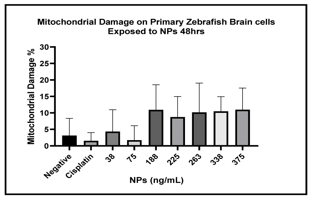
These results demonstrate that nZVI causes mitochondrial stress in a dose-dependent manner. This mitochondrial damage occurs without a corresponding apoptotic response, as shown in Figure 9 [13,14]. These results demonstrate that nZVI exposure induces significant mitochondrial membrane damage in primary zebrafish brain cells at concentrations lower than those required to elicit marked loss of viability. Importantly, this mitochondrial dysfunction occurs in the absence of statistically significant apoptotic activation, indicating that classical apoptosis is not the primary mechanism underlying nZVI-induced cytotoxicity in this mode [3,13,31]. The early onset of mitochondrial membrane damage at concentrations below the GI50 supports the notion that mitochondrial stress represents a primary cellular target of nZVI exposure [13,14]. Mitochondrial dysfunction can lead to energy failure, redox imbalance, and impaired cellular homeostasis without immediate activation of apoptotic pathways [3,12,13]. These findings provide a mechanistic explanation for the apparent discrepancy between viability loss and limited apoptotic activation observed in this study. Mitochondrial impairment is a well-recognized early indicator of iron-associated cellular stress and may precede multiple forms of regulated or non-regulated cell death [3,13,33,34]. Therefore, the observed effects are best interpreted as evidence of mitochondrial stress and iron-mediated cytotoxicity rather than definitive activation of a specific regulated cell death pathway. Although some apoptosis induction was observed, the one-way ANOVA with multiple comparisons analysis determined a non-statistically significant difference in comparison with the negative control (cells and DMSO). The dissociation observed between mitochondrial damage and apoptotic activation represents a key finding of this study. The reduction in PrestoBlue signal observed following nZVI exposure indicates a decrease in cellular metabolic activity under the experimental conditions. Because PrestoBlue is based on metabolic reduction, these findings should not be interpreted exclusively as a direct measurement of cell proliferation or cell death. The mitochondrial toxicity results provide complementary evidence of cellular dysfunction. However, the relationship between these endpoints cannot establish a temporal sequence of events because the measurements were evaluated following the same exposure period.
Similar patterns have been reported for iron-based nanomaterials, where mitochondrial dysfunction and metabolic collapse occur independently of classical apoptosis, particularly under conditions of iron overload or oxidative imbalanc e [3,13,31]. In such contexts, mitochondrial injury may reflect early cellular stress responses that precede or coexist with non-apoptotic modes of cell death [3,13,32].
Although ferroptosis has been increasingly recognized as an iron-dependent form of regulated cell death, confirmation of this pathway requires direct measurement of canonical markers such as lipid peroxidation and GPX4 activity [29-31]. In the absence of such measurements, ferroptosis is discussed here as a biologically plausible hypothesis rather than a demonstrated mechanism. Future studies incorporating these endpoints, as well as pharmacological modulation using pathway-specific inhibitors, will be necessary to delineate the molecular mechanisms linking nZVI exposure to mitochondrial dysfunction and loss of cellular viability. Taken together, the combined analysis of viability, apoptosis, and mitochondrial integrity reveals a consistent mechanistic pattern in which mitochondrial dysfunction emerges as an early and central event in nZVI-induced cytotoxicity. The absence of significant apoptotic activation despite reduced viability underscores the importance of non-apoptotic mechanisms in iron nanoparticle neurotoxicity. This observation aligns with previous studies reporting mitochondrial-driven toxicity and metabolic failure as primary responses to iron overload and nanoparticle exposure [3,13,31]. This study relies on an in vitro exposure model, which cannot fully replicate the complexity of in vivo systems, including blood-brain barrier dynamics, systemic metabolism, and immune responses [18,19].
Several limitations should be considered when interpreting these findings. Nanoparticle aggregation, sedimentation, and colloidal stability were not directly characterized in the cell exposure medium. Therefore, the nominal exposure concentrations may not fully represent the amount of nanoparticle material reaching the cultured cells. In addition, dissolved iron was not independently quantified during exposure, preventing a definitive distinction between effects associated with particulate nZVI and those potentially related to released iron species. These factors should be addressed in future studies to improve the characterization of nanoparticle exposure and the interpretation of neurotoxic responses. Additionally, the exposure concentrations employed represent an acute scenario and may not reflect environmentally relevant or chronic exposure conditions. These limitations are inherent to in vitro toxicology models and should be considered when extrapolating results to whole organisms.
Conclusion
This study employed an in vitro exposure model using primary brain cell cultures derived from adult zebrafish (Danio rerio) to investigate the neurotoxic potential of newly synthesized zero-valent iron nanoparticles. Direct exposure to nZVI resulted in a concentration-dependent reduction in cell viability, with limited activation of apoptotic pathways even at cytotoxic concentrations. In contrast, mitochondrial membrane integrity assays revealed significant mitochondrial dysfunction at lower concentrations than those associated with overt loss of viability, indicating that mitochondrial stress represents a key early cellular response to nZVI exposure. The observed dissociation between mitochondrial damage and apoptotic activation suggests that nZVI-induced cytotoxicity in this model is not primarily mediated by classical apoptosis, but rather involves mitochondrial impairment and iron-associated cellular stress mechanisms. While processes such as oxidative damage or iron-dependent regulated cell death pathways may contribute to these effects, additional targeted studies are required to define the precise molecular mechanisms involved. Although the direct exposure conditions used here do not replicate the complexity of in vivo systems, this primary neural cell model provides valuable mechanistic insight into the neurotoxic responses elicited by iron-based nanomaterials. These findings support the utility of zebrafish-derived neural cultures as a screening platform for evaluating potential neurotoxicity and informing the safe development and application of iron-containing nanoparticles.
Acknowledgements
The authors acknowledge the Institutional Development Award (IDeA) from the National Institute of General Medical Sciences of the National Institutes of Health, National Institutes of Health: grant number P20 GM103475-21.
Conflicts of interest
The authors declare no conflicts of interest regarding the publication of this paper.
- Tchounwou PB, Yedjou CG, Patlolla AK, Sutton DJ. Heavy metal toxicity and the environment. Experientia Suppl. 2012;101:133-164. Available from: https://doi.org/10.1007/978-3-7643-8340-4_6
- Jaishankar M, Tseten T, Anbalagan N, Mathew BB, Beeregowda KN. Toxicity, mechanism and health effects of some heavy metals. Interdiscip Toxicol. 2014;7(2):60-72. Available from: https://doi.org/10.2478/intox-2014-0009
- Valko M, Morris H, Cronin MTD. Metals, toxicity and oxidative stress. Curr Med Chem. 2005;12(10):1161-1208. Available from: https://doi.org/10.2174/0929867053764635
- Ward RJ, Zucca FA, Duyn JH, Crichton RR, Zecca L. The role of iron in brain ageing and neurodegenerative disorders. Lancet Neurol. 2014;13(10):1045-1060. Available from: https://doi.org/10.1016/S1474-4422(14)70117-6
- Liu M, Chen G, Xu L, He Z, Ye Y. Environmental remediation approaches by nanoscale zero-valent iron (nZVI) based on its reductivity: a review. RSC Adv. 2024;14:21118-21138. Available from: https://doi.org/10.1039/D4RA02789B
- Galdames A, Contreras D, Alarcón L, et al. Zero-valent iron nanoparticles for soil and groundwater remediation: recent advances. Int J Environ Res Public Health. 2020;17(16):5817. Available from: https://doi.org/10.3390/ijerph17165817
- Montiel Schneider MG, Martín MJ, Otarola J, et al. Biomedical applications of iron oxide nanoparticles: current insights, progress, and perspectives. Pharmaceutics. 2022;14(1):204. Available from: https://doi.org/10.3390/pharmaceutics14010204
- Ghazi R, Ibrahim TK, Nasir JA, et al. Iron oxide-based magnetic nanoparticles for hyperthermia, MRI, and drug delivery applications: a review. RSC Adv. 2025;15:11587-11616. Available from: https://doi.org/10.1039/D5RA00728C
- Pasinszki T, Krebsz M. Synthesis and application of zero-valent iron nanoparticles: environmental and biological implications. Nanomaterials (Basel). 2020;10(5):917. Available from: https://doi.org/10.3390/nano10050917
- Monopoli MP, Åberg C, Salvati A, Dawson KA. Biomolecular coronas provide the biological identity of nanosized materials. Nat Nanotechnol. 2012;7(12):779-786. Available from: https://doi.org/10.1038/nnano.2012.207
- Tran T, Nguyen M, Lin C, et al. Fate, transport, and toxicity of nanoparticles in biological and environmental systems. Sci Total Environ. 2023;912:169331. Available from: https://doi.org/10.1016/j.scitotenv.2023.169331
- Valko M, Jomova K, Rhodes CJ, Kuča K, Musílek K. Redox- and non-redox-metal-induced formation of free radicals and their role in human disease. Arch Toxicol. 2016;90(1):1-37. Available from: https://doi.org/10.1007/s00204-015-1579-5
- Semerad J, Navarro Pacheco NI, Grasserova A, et al. In vitro study of the toxicity mechanisms of nanoscale zero-valent iron (nZVI) and released iron ions using earthworm cells. Nanomaterials (Basel). 2020;10(11):2189. Available from: https://doi.org/10.3390/nano10112189
- Zeng G, He Y, Wang F, et al. Toxicity of nanoscale zero-valent iron to soil microorganisms and related defense mechanisms: a review. Toxics. 2023;11(6):514. Available from: https://doi.org/10.3390/toxics11060514
- Kirthi AV, Kumar G, Pant G, et al. Toxicity of nanoscaled zero-valent iron particles on tilapia (Oreochromis mossambicus). ACS Omega. 2022;7(51):47869-47879. Available from: https://doi.org/10.1021/acsomega.2c05696
- Rico CM, Majumdar S, Duarte-Gardea M, Peralta-Videa JR, Gardea-Torresdey JL. Interactions of nanoparticles with edible plants and their possible implications in the food chain. J Agric Food Chem. 2011;59(8):3485-3498. Available from: https://doi.org/10.1021/jf104517s
- Howe K, Clark MD, Torroja CF, et al. The zebrafish reference genome sequence and its relationship to the human genome. Nature. 2013;496(7446):498-503. Available from: https://doi.org/10.1038/nature12111
- Cassar S, Adatto I, Freeman JL, et al. Use of zebrafish in drug discovery toxicology. Chem Res Toxicol. 2019;32(1):62-74. Available from: https://doi.org/10.1021/acs.chemrestox.9b00335
- Fitzgerald JA, Jørs E, Suhr J, et al. Approaches to test the neurotoxicity of environmental chemicals in zebrafish. Environ Toxicol Chem. 2021;40(11):2834-2857. Available from: https://doi.org/10.1002/etc.4951
- Chen Z, Hendricks M, Cornelison K, et al. Primary neuron culture for nerve growth and axon guidance assays in zebrafish. PLoS One. 2013;8(3). Available from: https://doi.org/10.1371/journal.pone.0057539
- Odierna GL, et al. Generation of stable brain cell cultures from embryonic zebrafish. J Neurosci Methods. 2025:110255. Available from: https://doi.org/10.1016/j.jneumeth.2025.110255
- Adhish M, Manjubala I. Zebrafish disease models. Heliyon. 2023;9. Available from: https://doi.org/10.1016/j.heliyon.2023.e14557
- Wang CB, Zhang WX. Synthesizing nanoscale iron particles for rapid and complete dechlorination of TCE and PCBs. Environ Sci Technol. 1997;31(7):2154-2156. Available from: https://doi.org/10.1021/es970039c
- Phenrat T, Saleh N, Sirk K, Tilton RD, Lowry GV. Aggregation and sedimentation of aqueous nanoscale zero-valent iron dispersions. Environ Sci Technol. 2007;41(1):284-290. Available from: https://doi.org/10.1021/es061349a
- Li XQ, Elliott DW, Zhang WX. Zero-valent iron nanoparticles for abatement of environmental pollutants: materials and engineering aspects. Crit Rev Solid State Mater Sci. 2006;31(4):111-122. Available from: https://doi.org/10.1080/10408430601057611
- Nurmi JT, Tratnyek PG, Sarathy V, et al. Characterization and properties of metallic iron nanoparticles: spectroscopy, electrochemistry, and kinetics. Environ Sci Technol. 2005;39(5):1221-1230. Available from: https://doi.org/10.1021/es049190u
- Sun YP, Li XQ, Cao J, Zhang WX, Wang HP. Characterization of zero-valent iron nanoparticles. Adv Colloid Interface Sci. 2006;120(1-3):47-56. Available from: https://doi.org/10.1016/j.cis.2006.03.001
- Walkey CD, Chan WCW. Understanding and controlling the interaction of nanomaterials with proteins in a physiological environment. Chem Soc Rev. 2012;41(7):2780-2799. Available from: https://doi.org/10.1039/C1CS15233E
- Huang KJ, Wei YH, Chiu YC, Wu SR, Shieh DB. Assessment of zero-valent iron-based nanotherapeutics for ferroptosis induction and resensitization strategy in cancer cells. Biomater Sci. 2019;7(4):1311-1322. Available from: https://doi.org/10.1039/C8BM01525B
- Wu C, Shen Z, Lu Y, Sun F, Shi H. p53 promotes ferroptosis in macrophages treated with Fe3O4 nanoparticles. ACS Appl Mater Interfaces. 2022;14(38):42791-42803. Available from: https://doi.org/10.1021/acsami.2c10877
- Xuan L, Liu C, Zhang X, et al. Nanoparticles-induced potential toxicity on human health: mechanisms and evaluation. J Nanobiotechnology. 2023;21:257. Available from: https://doi.org/10.1186/s12951-023-01941-5
- Lomphithak T, Fadeel B. Cell death mechanisms and their implications in nanotoxicology. Toxicol Sci. 2023;192(2):141-160. Available from: https://doi.org/10.1093/toxsci/kfad041
- Liu S, Cheng W, Chen X, Zou M, Guo W, Feng X. Evaluating the safety and efficiency of nanomaterials: a focus on mitochondrial health. Biomed Pharmacother. 2024;180:117484. https://doi.org/10.1016/j.biopha.2024.117484
- Brand MD, Nicholls DG. Assessing mitochondrial dysfunction in cells. Biochem J. 2011;435(2):297-312. Available from: https://doi.org/10.1042/BJ20110162


 Save to Mendeley
Article Alerts
Subscribe to our articles alerts and stay tuned.
 This work is licensed under a Creative Commons Attribution 4.0 International License.